Western Blot (WB)—or immunoblot—is a workhorse immunoassay for many labs used to show antibody specificity, affirm gene expression, detect post-translational modifications (PTMs), diagnose ailments, and extra. Specific detection of bands akin to the protein of curiosity consequence from successively probing your blot with major and conjugated secondary antibodies. It is a extensively used methodology for detection of a selected protein in a posh matrix, comparable to cell or tissue lysate (i.e. protein extracts).
The Western blot assay makes use of gel electrophoresis (SDS-PAGE or native PAGE) to separate proteins in accordance with molecular weight. The proteins are then transferred from the gel onto a membrane (sometimes nitrocellulose or PVDF), which is then blocked with a protein blocking buffer to forestall non-specific binding and probed utilizing a major antibody to detect the protein of curiosity. This is adopted by an incubation with a secondary antibody conjugated to a reporter molecule, permitting the visualization of the goal protein; nonetheless, major antibodies conjugated to a reporter don’t require secondary antibody utilization. Wash steps utilizing a gentle detergent that accommodates buffer are additionally sometimes carried out after antibody incubations to take away any non-specific binding.
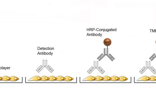
Western blot experiments will be carried out in a number of codecs, most of which require a conjugated secondary antibody to behave because the reporter molecule. Reporter molecules embrace horseradish peroxidase alkaline phosphatase enzymes, and fluorophores. When reporter enzymes are used, chromogenic, or luminescent substrates will be utilized for detection.
Fluorophore reporter molecules don’t require substrate however they do require specialised tools for knowledge assortment. Fluorescent detection is appropriate for multiplex WB experiments the place a number of targets will be detected in the identical assay utilizing fluorophore conjugates with non-overlapping emission spectra. Fluorescent WB can also be excellent for quantitative evaluation since detection permits for large dynamic ranges and sign normalization.
The selection of WB membrane will depend on the kind of experiment to be carried out. Most widespread membranes used are nitrocellulose or polyvinyldifluoride (PVDF). Nitrocellulose is straightforward to make use of and supplies appropriate knowledge for most typical enzymatic reporter experiments. Low fluorescent PVDF membranes are really helpful for fluorescent Western blot functions.
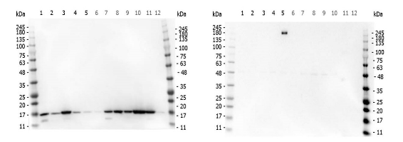
Multi-lysate WB
As application-specific tips and requirements for validating analysis antibodies more and more turns into a topic of scrutiny by the scientific neighborhood, a number of approaches can—and will—be carried out to show the specificity of a major antibody with enough robustness. One such method is the affirmation of target-specific antibody properties by the inclusion of multi-lysate panels from cells recognized to specific or not the goal of curiosity based mostly on genomic and proteomic research. The acceptable destructive controls for cells naturally producing the goal are the identical cells wherein the abundance of the goal is selectively altered by chemically stimulation or genetic approaches.
Rockland routinely scrutinize the specificity or its major antibodies by assessing their efficiency in multi-lysate Western blots. Quite a lot of situations are evaluated for every goal in order that specificity, sensitivity and reproducibility will be decided. This ensures dependable efficiency and make sure lot-to-lot consistency.
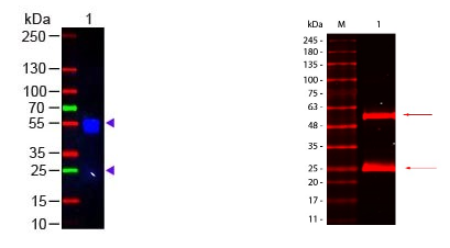
Immunoprecipitation-WB
Immunoprecipitation (IP) is likely one of the most generally used approaches for antigen purification and detection. The method makes use of antigen-specific antibodies to isolate an antigen of curiosity from a posh protein combination that’s subsequently analyzed by Western blotting in an effort to assess the relative quantity and measurement of the goal antigen itself and/or target-associated proteins.
Analysis of immunoprecipitated proteins by immunoblotting will be difficult as a result of the reagent used to detect the WB staining antibody will usually bind to the heavy and light-weight chains of the precipitating antibodies. As described beneath, this downside will be simply corrected through the use of our TrueBlot® merchandise that selectively bind staining antibodies solely by elevated sensitivity, much less background noise, and enhanced accuracy.
IP Western blots present extremely particular outcomes, but usually endure from heavy/mild chain blotting, contamination, and ongoing interference. TrueBlot® merchandise clear up almost all of those issues by elevated sensitivity, much less background noise, and enhanced accuracy. TrueBlot® reagents allow you to generate clear, best-quality knowledge in your Immunoprecipitation and Western blot protocols. Available in a number of choices, from IP Beads alone, to full IP/Western blot kits from goat, mouse, rabbit, or sheep.
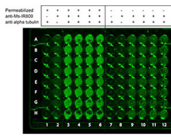
In-Cell Western
In-cell western (ICW) assays—or cell based mostly assays, cell-based ELISA, In-Cell ELISA (ICE), or Fast Activated Cell-based (FACE) ELISA—permit researchers a easy and fast assay methodology for the quantification biomarkers and signaling proteins in complete cells utilizing antibodies. The Odyssey® infrared imaging system by LICOR® is nicely fitted to ICW; nonetheless, different imaging methods may additionally be used. The methodology combines the specificity of a WB with the quantification of an ELISA. ICW assays can be utilized for detection of proteins in fastened cells or proteins with physiological- or biologically-relevant mobile context and in multiplex quantification of two targets utilizing 700 nm and 800 nm channels with acceptable dye-conjugated antibodies.
Many of Rockland’s antibodies have been validated to be used in ICW and since ICW assays are associated to immunofluorescence (IF), most antibodies which have been validated and accepted for IF will be equally used with ICW assays as nicely.
Secondary Antibodies for WB
Secondary antibody conjugates are perfect for Western blotting. When selecting a secondary antibody conjugate for an antibody assay consideration should be given to focus on species, conjugate (i.e. peroxidase, FITC, Biotin) and host species. In addition to plain secondary antibodies, Rockland gives pre-adsorbed secondary antibodies that are appropriate for detection strategies the place cross-reactivity could also be a difficulty.
Reporter enzymes are used extensively in molecular biology as a result of they permit visualization or detection of immune complexes. Horseradish Peroxidase (HRP) is a extensively used reporter enzyme, and relying on the substrate it will possibly yield a chromogenic, or luminescent product (chemiluminescence). Alkaline phosphatase can also be used, most sometimes because the reporter in chromogenic Western blot assay format.
Antibodies Conjugated to Alkaline Phosphatase (AP or Alk Phos) are used within the detection of proteins in Western blotting and ELISA immunoassay procedures. The alkaline phosphatase (AP) catalyzes colorimetric reactions utilizing BCIP/NBT Substrates or FemtoMax chemiluminescent substrate. Secondary Antibody conjugates are conjugated to the best grade of alkaline phosphatase utilizing Rockland’s proprietary know-how.
Rockland conjugates a broad group of secondary antibodies to lots of the basic and subsequent technology of fluorescent markers together with fluorescein, Texas Red, Phycoerythrin. Rockland additionally produces many subsequent technology flurochrome dyes. These are designed for detection of major antibodies in multiplex, multi-color evaluation. Next technology fluorochrome conjugates (Atto-tec dyes, DyLight™ dyes) provide superior absorption (excessive extinction coefficient), excessive fluorescence quantum yield and superior excessive photostability. All of the conjugates are perfect for varied immunofluorescence based mostly assays together with fluorescent Western blotting, immunofluorescence microscopy, FLISA, and extra.
Substrates for WB
Chromogenic Substrates
Chromogenic substratesare utilized in colorimetric assays since they lead to a measurable shade change within the presence of an enzyme-antibody complicated certain to particular analytes. For WB utilizing horseradish peroxidase (HRP) in colorimetric detection TMB and DAB substrates are generally used. Alkaline phosphatase (AP) chromogenic substrates embrace BCIP/NBT, which often exhibit the best sensitivity and dependable detection of AP exercise.Chromogenic blotting substrates can be found from Rockland in a wide range of specs and codecs. The acceptable substrate selection will depend on the enzyme label, desired sensitivity and type of sign or methodology of detection wanted.
The peroxidase response with our TMBM substrate produces a water-soluble blue product that may be precipitated onto a membrane. The precipitating product produces blue to darkish blue bands within the enzyme location. TMBM is nicely suited to functions that require excessive signal-to-noise. DAB is one other peroxidase substrate and yields a brown precipitate within the presence of HRP and peroxide.
The NBT/BCIP reagent can also be generally utilized in chromogenic Western blot immunoassays. NBT serves as an oxidant and BCIP because the alkaline phosphatase substrate. Together NBT and BCIP type reactants within the presence of alkaline phosphatase which yields a darkish purple to black, water-insoluble, precipitant product offering robust sensitivity.
Chemiluminescent Substrates
Rockland produces a number of luminol based mostly substrates with chemiluminescence for the detection of horseradish peroxidase (HRP). PicoMax™ and FemtoMax™ are designed for top efficiency in Western blotting and are practical on each nitrocellulose and PVDF membranes. FemtoMax™ produces chemiluminescence and permits for the detection of all the way down to femtogram (10-15) quantities of antigen. Detection strategies might embrace photographic movie or different imaging strategies, together with extremely delicate CCD digicam based mostly methods.
within the different hand provide a number of benefits over the chromogenic substrates. Mainly, these methods are considerably extra delicate for detection of enzymatic exercise with out using radioactive isotopes, luminescent detection sometimes occurs inside couple of minutes and the sign is extra amenable to quantification as a result of it requires using digital charge-coupled gadgets (CCD) for detection that permit for a large dynamic vary utilizing extended publicity.
Blocking Buffers for WB
When performing a Western blot, the blocking buffer shouldn’t be ignored. The blocking buffer fills within the places on the membrane that may bind protein and trigger background if not handled. The important reagent in a blocking buffer is protein, the place the protein is non-antibody reactive. Popular blocking proteins embrace non-fat dried milk (NFDM), BSA, casein, and mixtures thereof. Of observe, a single blocking agent will not be enough for all Western functions. Some blocking brokers can intervene with major antibody exercise, could also be incompatible with the reporter system in use, or produce undesired auto-fluorescence. We develop a number of blocking buffer reagents appropriate for all Western blot functions, together with BLOTTO-NFDM and BSA for traditional functions, and a specifically formulated blocking buffer for fluorescent Western blotting.
Fluorescent blocking buffers are specifically formulated to protect the fluorescence from fluorochrome-dye conjugated secondary antibodies and likewise present an ultra-low background in comparison with different blocking buffers. Our buffers work with many fluorescent secondary antibody conjugates together with, FITC, IRDye®, Atto dye, and DyLight dye. Our Blocking Buffer for Fluorescent Western Blotting is exceptionally good in commonplace chemiluminescent blotting functions and is demonstrated to be superior for 2D WB experiments in head-to-head comparisons with different blocking reagents.
Kits for WB
Western blot kits could also be obtainable in your assay, simplifying your reagent wants. We provide kits for chemiluminescence, fluorescent, and chromogenic immunoassay codecs. Our kits are configured with easy and easy-to-use protocols for each newbie and knowledgeable customers alike. Kits are species-specific for detection of mouse or rabbit major antibodies and are available prepared as a format-specific bundle, together with a membrane blocking reagent, washing buffers, secondary antibodies, and substrate (if required). Some of our extra widespread kits embrace FentoMax kits for chemiluminescent functions, infrared (IR) and Dylight™ kits for detection on fluorescent Western blot protocol imaging methods.